- Gaseous Exchange in Mammals
- Air path: nose/mouth (filtered, moistened by mucus) → pharynx → past the epiglottis (covers the glottis to stop food entering the trachea) → larynx → trachea (held open by cartilage rings, lined with ciliated mucus membrane) → bronchi → bronchioles → alveoli.
- The trachea and bronchi have smooth muscle and cartilage (for support); the smallest bronchioles lack cartilage and can constrict fully. 12 pairs of ribs form a protective cage; pleural membranes (with lubricating pleural fluid) line the thoracic cavity.
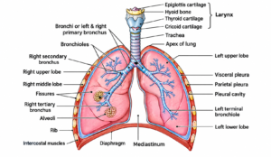
Structure of the human respiratory tract and lungs
Structure and Adaptation of the Alveoli
- ~7 million alveoli give a surface area of 100–150 m²; walls are one cell thick (squamous epithelium), moist, permeable, and richly supplied with capillaries from the pulmonary artery.
- Surfactant: a detergent-like substance that lowers surface tension (so alveoli don’t collapse and less effort is needed to inflate the lungs), speeds gas transport, and kills bacteria.